Dec 20, 2011 at 9:58 am. The process of setting the ball back into the socket after the arthrogram is known as a closed reduction.

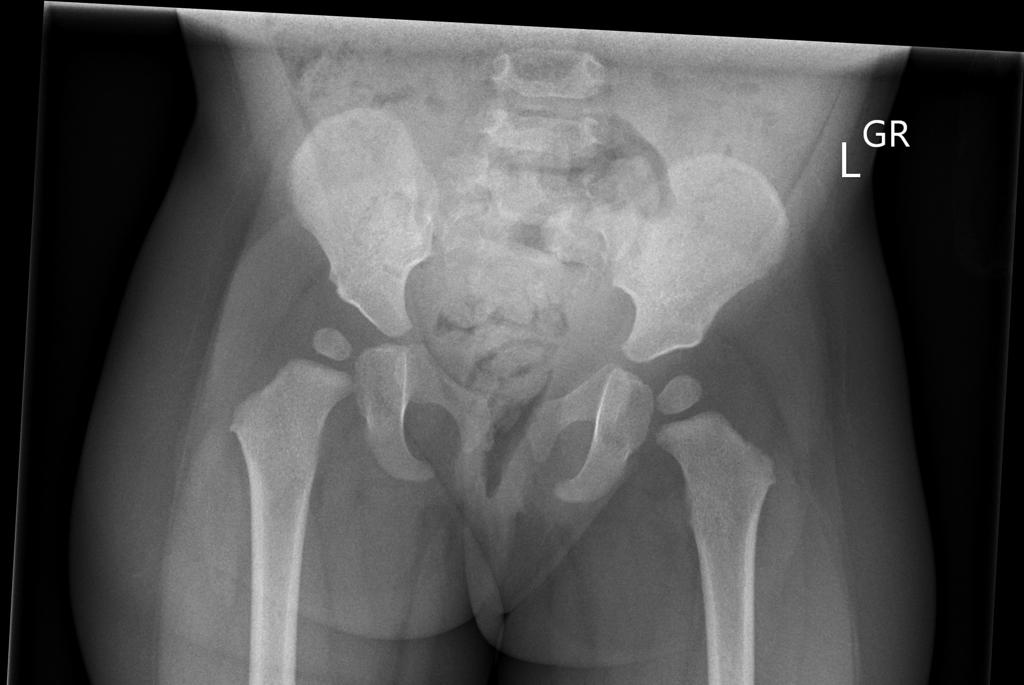
Pelvic X-ray - Different Ages Radiology Case Radiopaediaorg
This means that baby’s thighs are spread around the parent’s torso and baby's hips are open with his knees are bent at the same height.

How do they x ray babies hips. It has nothing to do with whether or not the parents will hold the infant. Hip ultrasounds are recommended for infants from birth to six months old because their bones have not fully developed. Radiographers appear to like it.
Two tests are performed, called the barlow and ortolani tests, to examine the function of the hip joints. It’s used because babies and toddlers are incapable of following directions to hold still. If she does have it they may try to brace it first.
In babies with hip dysplasia, the joint has not formed normally, and the hips are prone to moving in and out of joint. The most concerning physical exam finding is the classic ortolani sign — the hip is felt to “clunk” into place with abduction of the hip. Your baby should have an ultrasound scan of their hip between 4 and 6 weeks old if a doctor, midwife or nurse thinks their hip feels unstable.
This test is called an arthrogram. Those things work great, one person wrote on reddit. The examination involves gently moving your baby's hip joints to check if there are any problems.
The abscess is usually located in the metaphysis of long bones, but may be located in the epiphysis in young children. A barlow sign — when the hip slides out of the socket with posteriorly directed force — also indicates risk of ddh. Under anesthesia, the doctor will insert a very fine needle in the baby’s hip and inject contrast so they can clearly view the ball and the socket.
As the hips are moved in these tests, a. It should not cause them any discomfort. While the images are being taken, you’ll need to hold your breath and remain still to get the clearest.
Whichever style of baby carrier or sling you choose, make sure your baby’s hips are spread out in the squat position. This is necessary to make the diagnosis or to be sure the hip is normal. It is put on by an orthopedic surgeon while using x.
The hip joint can be imaged under various angles. It's a cast that goes around both hips and down the leg to keep the hips aligned. If it persists they may put on a spica cast.
2

Pin On Awesomeness
X-ray Screening - International Hip Dysplasia Institute

Developmental Dysplasia Of The Hip Ddh Developmental Dysplasia Of The Hip Pediatric Nursing Physical Therapy Assistant

Pin On X-ray And Medicine

Lower Limb Radiographs Anatomy And Physiology Anatomy Sacroiliac Joint

Orthoroentgenogram Radiology Imaging Body Anatomy Medical Imaging

Leerburg The Importance Of Good Positioning On Canine Hip X-rays Canine Hips Mastiffs

X-ray Of Baby Lilly Doll Xray Art Contemporary Graphic X Ray

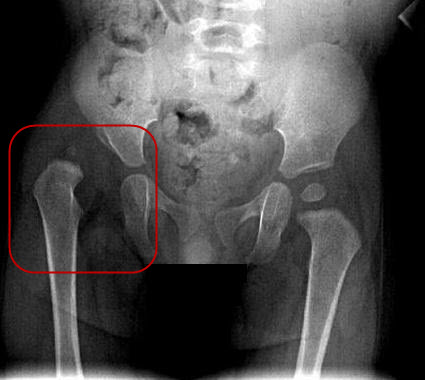
Pelvis X-ray Ap View Showing Left Sided Dysplastic Hip With Femur Download Scientific Diagram

Normal Pelvis X-ray - 9-year-old Radiology Case Radiopaediaorg
Developmental Dysplasia Of The Hip Radiology Case Radiopaediaorg

Pelvic X-ray - Different Ages Radiology Case Radiopaediaorg
Normal Pelvis X-ray - 4-year-old Radiology Case Radiopaediaorg

Developmental Dysplasia Of Hip Or Congenital Dislocation Of Hip Bone And Spine Radiology Developmental Dysplasia Of The Hip Radiology Imaging
Pin On Iyt Health Advisor

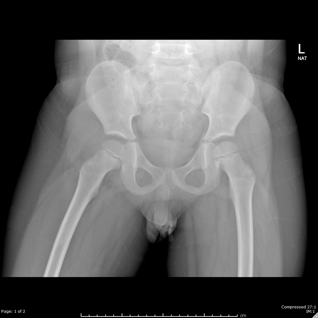
X-ray Pelvis With Both Hip Joints In A 10 Months Old Infant Showing A Download Scientific Diagram

This Is How They X-ray Babies Education Motivation Educational Technology X Ray

Interpreting X-rays Of The Pelvis Hip Joint And Femur - Youtube

